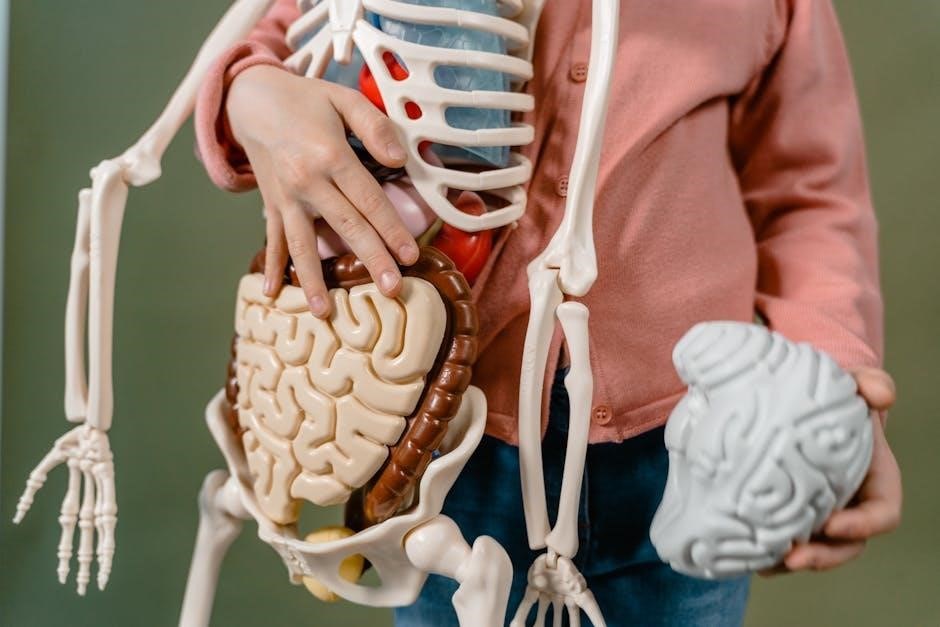
Netter’s Atlas of Human Anatomy is a cornerstone of medical education, offering detailed, physician-illustrated views of the human body. The 7th edition includes updated plates for clarity and accuracy, making it an essential resource for students and professionals alike.
Overview of the Atlas
Netter’s Atlas of Human Anatomy is a renowned medical resource that provides detailed, anatomically accurate illustrations of the human body. The 7th edition features updated plates, enhancing clarity and precision. Illustrated by physicians, it offers a clinician’s perspective, making it invaluable for both students and professionals. The atlas combines a regional and systemic approach, ensuring comprehensive coverage of anatomy. Its clear, brilliant detail and organized structure make it a cornerstone in medical education and practice, providing a visual foundation for understanding human anatomy.
Significance of the Atlas in Medical Education
Netter’s Atlas of Human Anatomy holds a pivotal role in medical education, serving as a visual cornerstone for understanding human anatomy. Its detailed, physician-illustrated plates provide unparalleled clarity, bridging the gap between theoretical knowledge and clinical practice. The 7th edition’s updated content ensures relevance, while its organized structure caters to diverse learning needs. The atlas is widely regarded for its ability to simplify complex anatomical concepts, making it an indispensable tool for students, educators, and healthcare professionals; Its integration into modern teaching tools further enhances its educational value, solidifying its place as a foundational resource in medical training.
Key Features of the 7th Edition
The 7th edition of Netter’s Atlas of Human Anatomy introduces updated illustrations, including 57 revised, 200 relabeled, and 17 entirely new plates, enhancing clarity and accuracy. The atlas retains its classic regional approach while incorporating a systemic perspective for comprehensive understanding. Digital features, such as the Interactive Dissector, offer labeled and unlabeled views, aiding interactive learning. The edition also includes surface anatomy and imaging plates, bridging anatomical knowledge with clinical practice. These updates ensure the atlas remains a vital resource for medical students and professionals, combining traditional excellence with modern educational tools.

Historical Background of the Atlas
First published in 1989, Netter’s Atlas of Human Anatomy was created by Frank H. Netter, whose detailed illustrations revolutionized medical education and remain unmatched today.
Frank H. Netter and His Contributions to Anatomy
Frank H. Netter, MD, was a visionary physician and artist whose detailed, accurate illustrations transformed medical education. His unique ability to combine clinical expertise with artistic precision made his work unparalleled. Netter’s Atlas of Human Anatomy, first published in 1989, became a cornerstone of anatomical studies. His illustrations are renowned for their clarity, depth, and precision, making complex anatomy accessible to students and professionals. Beyond the atlas, Netter contributed to numerous medical texts and the CIBA pharmaceutical series, leaving a lasting legacy in the field of anatomy and medical visualization.
Evolution of the Atlas Since Its First Publication
Since its first publication in 1989, Netter’s Atlas of Human Anatomy has undergone significant evolution, with each edition refining its content and presentation. The 4th edition introduced revised and relabeled plates, while the 7th edition featured updated illustrations and a digital version. The 8th edition expanded further, adding new plates and enhancing clarity. These updates have maintained the atlas’s reputation for precision and accessibility, ensuring its continued relevance in medical education and practice. The integration of interactive and digital versions has also catered to modern learning preferences, solidifying Netter’s legacy as a cornerstone of anatomical study.
Impact of the Atlas on Medical Illustration
Netter’s Atlas of Human Anatomy revolutionized medical illustration by setting a new standard for clarity and precision. Frank H. Netter’s distinctive artwork, combining anatomical accuracy with artistic elegance, influenced generations of medical illustrators. The atlas’s detailed, physician-perspective illustrations became a benchmark, shaping how anatomy is visually represented. Its success also spurred advancements in digital anatomy tools, integrating traditional art with modern technology. This fusion has made Netter’s Atlas a cornerstone in medical education, inspiring both traditional and digital anatomical resources. Its enduring influence underscores its role in bridging art and science in medical visualization.

Structure and Organization of the Atlas
Netter’s Atlas of Human Anatomy is organized regionally and systemically, offering detailed illustrations. The 7th edition includes updated plates, with both regional and systemic approaches, ensuring comprehensive coverage. Interactive digital versions enhance accessibility, making it a versatile tool for learners and professionals.
Regional Approach to Anatomy
Netter’s Atlas of Human Anatomy employs a regional approach, organizing content by body regions. This method allows for a logical and systematic study of anatomy, correlating structure with function.
The 7th edition features updated plates, enhancing clarity and detail. Regional dissections and cross-sectional views provide a comprehensive understanding, making it ideal for both students and professionals in clinical practice.
Systemic Approach to Anatomy
Netter’s Atlas of Human Anatomy also incorporates a systemic approach, focusing on individual organ systems. This method allows for a detailed understanding of each system’s structure and function, aiding in clinical correlation. The 7th edition includes updated illustrations, enhancing the clarity of systemic anatomy. This approach complements the regional method, providing a comprehensive learning experience for medical students and professionals. The systemic perspective is particularly useful for understanding complex anatomical relationships and their relevance to medical practice.
Interactive and Digital Versions of the Atlas

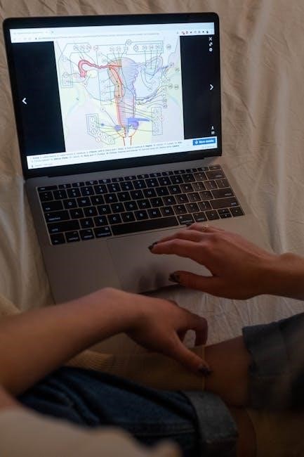
Netter’s Atlas of Human Anatomy is available in interactive and digital formats, enhancing learning through innovative tools. The 7th edition offers digital features like labeled and unlabeled plates, allowing users to test their knowledge. Interactive dissectors provide a three-dimensional view, enabling deeper exploration of anatomical structures. These digital versions are accessible on devices like iPads, making them ideal for modern medical education. They cater to diverse learning styles, offering a flexible and engaging way to master human anatomy. This integration of technology ensures the atlas remains a cutting-edge resource for both students and professionals in the medical field.
Medical Applications of the Atlas
Netter’s Atlas of Human Anatomy is widely used in medical education and clinical practice, serving as an essential reference for understanding human anatomy in detail.
Use in Medical Schools and Training Programs

Netter’s Atlas of Human Anatomy is a cornerstone in medical education, widely used in schools and training programs worldwide. Its clear, detailed illustrations provide students with a comprehensive understanding of human anatomy. The atlas is particularly valued for its physician-illustrated plates, which offer a clinical perspective essential for future professionals. Updated editions include new plates and labels, enhancing learning experiences. Many institutions integrate the atlas into their curricula, while students rely on it for self-study and exam preparation. Its clarity and accuracy make it an indispensable tool for mastering anatomy, a foundational skill in medical training.
Application in Clinical Practice

Netter’s Atlas of Human Anatomy is widely used in clinical practice by surgeons, radiologists, and other healthcare professionals. Its detailed, accurate illustrations serve as a trusted reference for understanding complex anatomical structures. Clinicians rely on the atlas for preoperative planning, intraoperative guidance, and educating patients. The clear, physician-illustrated plates provide a practical perspective, aiding in diagnosis and treatment decisions. Updated editions incorporate modern imaging techniques, aligning with advancements in medical practice. This makes the atlas an invaluable resource for bridging anatomical knowledge with real-world clinical applications, ensuring precise and informed patient care.
Integration with Modern Teaching Tools
Netter’s Atlas of Human Anatomy seamlessly integrates with modern teaching tools, enhancing learning experiences for medical students and professionals. The atlas is available in digital formats, including interactive dissectors, allowing learners to explore anatomy dynamically. It complements virtual dissection software and online platforms, providing a comprehensive educational experience. Institutions often pair the atlas with iPads and other digital devices for hands-on learning. Additionally, the atlas aligns with online resources and teaching modules, making it a versatile tool for both traditional and modern pedagogical approaches. This integration ensures that anatomical knowledge remains accessible and engaging in contemporary medical education.
Accessing the Netter Atlas of Human Anatomy PDF
The Netter Atlas of Human Anatomy PDF is widely available for download from various sources, including the official Elsevier website and academic platforms. It offers digital access to detailed anatomical illustrations, making it a convenient resource for medical students and professionals;
Downloading the PDF Version
Downloading the Netter Atlas of Human Anatomy PDF is straightforward, with options available from the official Elsevier website, Google Drive, and other academic platforms. The 7th and 8th editions are widely accessible, offering high-resolution illustrations. Files range from 1.0GB to 100.6MB, ensuring compatibility with various devices. Users can access labeled or unlabeled versions, depending on their preference. The PDF format allows for interactive features, such as zooming and searching, enhancing its utility for study and reference. Ensure to download from reputable sources to maintain quality and authenticity of the content.
Legality and Ethical Considerations of Sharing the PDF

Sharing the Netter Atlas of Human Anatomy PDF without proper authorization may infringe on copyright laws, as the content is owned by Elsevier. Distribution without permission violates intellectual property rights and can lead to legal consequences. Ethically, it is important to respect the work of authors and publishers by purchasing or accessing the PDF through legitimate channels. Institutions and individuals should adhere to copyright guidelines to support the creation of high-quality educational materials. Always ensure that access to the Atlas is obtained legally and ethically to uphold academic integrity.
Supplementary Resources and Guides
Netter’s Atlas of Human Anatomy is complemented by various supplementary resources to enhance learning. These include interactive digital tools, such as the Interactive Dissector, which allows users to explore anatomy in depth. Study guides and flashcards are also available to aid in memorization. Additionally, the atlas is supported by online platforms that offer labeled and unlabeled plates for self-testing. These resources are designed to cater to both students and professionals, providing a comprehensive learning experience. They integrate seamlessly with the atlas, ensuring a well-rounded understanding of human anatomy.

Comparative Analysis with Other Anatomy Resources
Netter’s Atlas is renowned for its physician-illustrated, clinically focused views, distinguishing it from resources like Gray’s Anatomy, which offers more detailed textual descriptions alongside visuals.
Netter vs. Gray’s Anatomy
Netter’s Atlas and Gray’s Anatomy are two iconic anatomy resources. Netter’s Atlas is celebrated for its clinically oriented, physician-illustrated visuals, emphasizing clarity and practicality. In contrast, Gray’s Anatomy provides exhaustive textual descriptions alongside detailed illustrations, catering to both students and professionals. While Netter’s focus is on concise, visually driven learning, Gray’s offers a more comprehensive, traditional anatomical reference. Both are indispensable, but Netter’s is often preferred for its straightforward, clinically relevant approach, making it a favorite among medical students and practitioners seeking quick, precise anatomical insights.

Netter vs. other Anatomical Atlases
Netter’s Atlas stands out among anatomical atlases for its unique combination of artistic detail and clinical relevance. Unlike others, it is illustrated by physicians, ensuring accuracy and a focus on structures most pertinent to medical practice. While other atlases may emphasize complexity or exhaustive detail, Netter’s clarity and concise presentation make it a preferred choice for students and professionals. Its regional and systemic approaches, along with updated digital features, provide a versatile learning tool that surpasses many traditional anatomical resources in practicality and accessibility.
Unique Features of Netter’s Atlas
Netter’s Atlas of Human Anatomy is renowned for its exceptional illustrations, created by physicians, ensuring anatomical accuracy and clinical relevance. The 7th edition features updated plates, enhancing clarity and detail. Its regional and systemic approaches provide a comprehensive understanding of human anatomy. Unlike other atlases, Netter’s combines artistic precision with a focus on structures vital to medical practice. The inclusion of interactive digital versions and supplementary tools further distinguishes it, offering a versatile learning resource for both students and professionals. Its unique blend of tradition and innovation solidifies its position as a leading anatomical reference.
Netter’s Atlas of Human Anatomy remains a cornerstone in medical education, offering unparalleled clarity and detail. Its evolution over editions, particularly the 7th edition, ensures it stays relevant and indispensable. The unique combination of artistic precision and clinical focus makes it a standout resource. With the addition of digital versions, it adapts to modern learning needs. As a trusted reference, it continues to bridge the gap between anatomy and clinical practice, solidifying its legacy as an essential tool for students and professionals alike.
